 Use the AI Canvas.
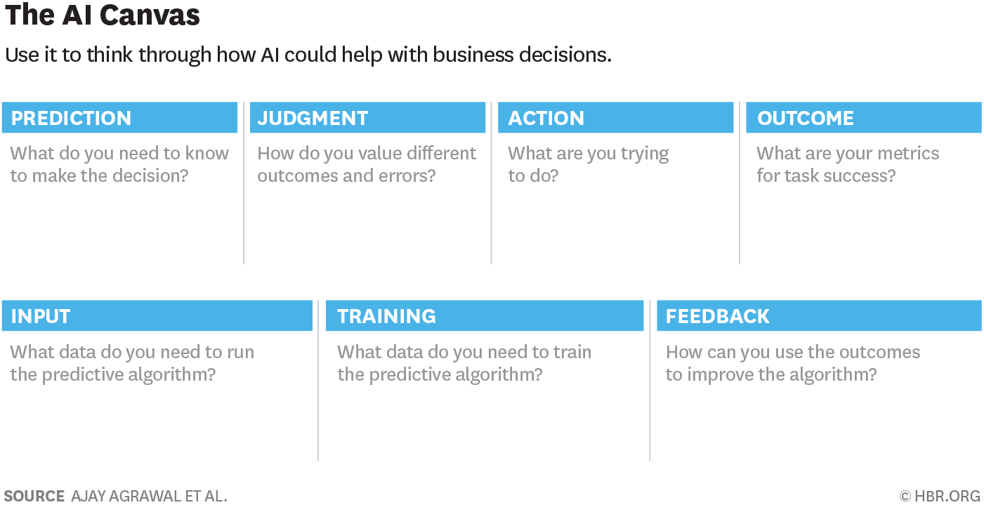
Source: A Simple Tool to Start Making Decisions with the Help of AI
In a recent Harvard Business Review article, Ajay Agrawal and coauthors shared a simple tool to think about how an AI tool may be deployed. Although the tool is no more complex than 6 boxes of free text, it does follow a number of best practices when thinking about general data and machine learning:
- Always define an end-goal – what’s the desired outcome?
- You should always make a hypothesis of what may drive this desired outcome.
- You should determine how to present the ML prediction in a way that drives action, not just the data itself.
- Your data acquisition strategy should include a feedback mechanism.
For example, this is how one might fill out the AI Canvas tool in a radiology use case:
- Prediction: Predict whether a brian MRI for a cancer patient contains increasing or new hydrocephalus
- Action: Label the examination as critical, and denote that AI has determined a critical finding. For example, create an “AI-STAT” category on worklist priority.
- Judgment: Compare the cost of interpreting this brain MRI at its usual turnaround time, versus immediately.
- Outcome: Observe whether the action taken in response to a study labeled AI-STAT is correct.
- Input: New MRIs of the brain MRI performed, and their prior studies.
- Training: Historical brain MRIs
- Feedback: Identify false positives – perhaps the prior study was from 20 years ago, or there’s been surgical resection, so that ex vacuo dilation of ventricles is not hydrocephalus. Perhaps there has been recent surgery Identify false negatives – subtle enlargement of the temporal horns missed by AI. Use this information to improve the AI.
How might you use this worksheet to brainstorm AI for your radiology practice?