Getting Started with NVidia Jetson Nano via JetBot

As part of the effort to explore NVidia Jetson Nano, and part of its AI Specialist course (after finishing Fundamentals of AI in Nvidia Deep Learning Institute), I started buliding a JetBot.

JetBots are well documented and relatively easy to build provdied you have the right parts. There is also a bill of materials to make purchasing simpler.

The chassis was 3D printed according to the full DIY instructions (did not use a kit).

The camera used in this picture is actually from a Rasp Pi infrared camera I bought years ago. Turns out I could remove the lens and apply to another camera I bought for this project (IMX290-160FOV). It turns out that the 70 degree FOV on the lens was really just not wide enough to see what is going on. The 160-degree FOV was perfect and seems to help the bot see around itself.

This post is part of a series on learning about Internet of Things. These posts are mainly a learning tool for me – taking notes, jotting down ideas, and tracking progress. This means they might be unrelated to radiology or healthcare. They also might contain works-in-progress or inaccuracies.

Different flavors of ESP8266

ESP8266 is a wifi enabled microcontroller. One of the most helpful ones because of it’s wifi ability and very low cost. This makes the ESP8266 popular in even commercial products that need wifi connectivity.

For development purposes, there are also a lot of variants for this chip. After some preliminary research, there appears to be two most helpful breakout boards for it.

NodeMCU

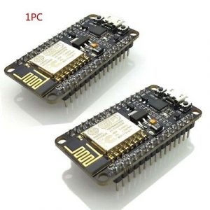
NodeMCU / ESP8266

NodeMCU is technically the name of the Lua-compatible firmware for ESP8266, which later added support for ESP32 (the more powerful, dual-core sibling of ESP8266). NodeMCU was created in 2014 when user Hong committed the first file of nodemcu-firmware to GitHub. but people sometimes use this term to refer to breakout boards using ESP8266 following this particular schema. It comes with additional chips that enable USB-to-serial and other “quality of life” enhancements that make development easier. The breakout board is also compatible with solderless breadboards, making prototyping much easier.

Continue reading

UT Audit of MD Anderson / IBM Collaboration

The details can be downloaded here.

A few snippets…

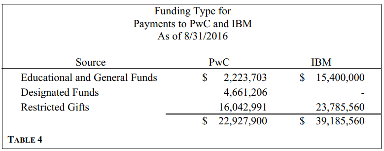
“Gift funds used in support of the OEA project have a deficit balance of $11.59 million as of August 31, 2016, meaning that MD Anderson spent gift monies it has not yet received from donors.”

“Agreement with PricewaterhouseCoopers (PwC) for “Business Plan for a Flagship Informatics Tool” to lead an assessment of the “capabilities necessary to build the tool” and “incorporate the outcome of the assessment into a business plan that will guide the development” of the tool.”

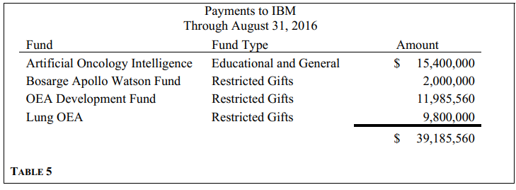
“The first MD Anderson contract related to development of OEA using Watson technology was signed with IBM in June 2012… The original contract terms were for six months at a fixed fee of $2.4 million. That contract has been extended 12 times, with total fees of $39.2 million. The current extension expired on October 31, 2016.”

Interestingly, if you search in the document for “radiology” or “imaging”:


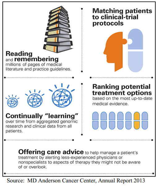
That said, it is a good cautionary tale for the radiologist-informaticist because the value proposition very closely mirrored what we are hearing in imaging today.

Just swap out the words “treatment,” “clinical-trial,” and “therapy” with “diagnosis” and “imaging”:

Beyond “Embrace or Perish” – Thrive in the AI Era By Thinking Different

Artificial intelligence promises to uproot the practice of medical imaging – traditionally thought to be expensive and highly expert-driven. Radiology industry juggernauts like General Electric, Nuance, and Partners HealthCare all teaming up with established AI players like Intel and NVidia. The innovations have advanced rapidly. Recently, AI has managed to achieve super-human accuracy in the detection of pneumonia on radiography.

More interestingly, some vendors are marketing FDA-approved AI to expedite stroke treatment and eliminating diagnostic delays. The definition of “delay?” Human radiologists taking time to read those head CTs.

So what’s a radiology practice to do?

What can we learn from other industries that have seen the arrival of large, untamable, data- and AI-powered competitors?

Amazon entering an industry is typically regarded as an extinction-level event. Amazon started with the Internet, then had Big Data, and now has AI – they’ve bet first, bet big, and bet right on all of the major tech trends.

But this story isn’t about Amazon; it’s about everybody else.

Continue reading

Your Radiology AI Briefing – January 3, 2019

In this briefing:

  • Research: F-18 FDG PET-CT predicts Alzheimer’s disease with 100% sensitivity and 82% specificity
  • Research: An “explainable AI” model for intracranial hemorrhage
  • Hardware: Specialized AI hardware promises to accelerate network training over 1000-fold in 5 years.
  • Startup: Philips funds 19 AI-related imaging startups in 14 countries
  • Marketing: Healthcare AI expected to grow 20-fold by 2024

Radiology AI Briefing logo graphic

Stay up to speed in 2 minutes. Radiology AI Briefing is a semi-regular series of blog posts featuring hand-picked news stories and summaries on machine learning and data science.


Continue reading

Your Radiology AI Briefing – May 12, 2018

In this briefing:

  • Research: Machine learning algorithm predicts wait time for outpatient imaging.
  • Commercial brain imaging AI receives FDA clearance
  • Paul Chang shares insight on the future of AI
  • Dreyer and Allen publish their views on the radiology AI ecosystem
  • CB Insights publishes market research on Google’s increasing involvement in healthcare AI.

Radiology AI Briefing logo graphic

Stay up to speed in 2 minutes. Radiology AI Briefing is a semi-regular series of blog posts featuring hand-picked news stories and summaries on machine learning and data science.


Continue reading

Your Radiology AI Briefing – May 3, 2018

In this briefing:

  • RSNA launches a new AI journal.
  • ACR makes several moves to advance the use of artificial intelligence in the future of medical imaging.
  • Researchers publish in high impact journal the successful use of AI to detect breast density.
  • ACR and MICCAI sign agreement to advance AI in medical imaging.
  • Geisinger declares successful implementation of algorithm to improve time-to-diagnosis of intracranial hemorrhages 20-fold.

Radiology AI Briefing logo graphic

Stay up to speed in 2 minutes. Radiology AI Briefing is a semi-regular series of blog posts featuring hand-picked news stories and summaries on machine learning and data science.


Continue reading

What’s in Store for Our VA Health System?

Source: https://www.philadelphia.va.gov

Several days ago, Rear Adm. Ronny Jackson confirmed he will drop out of the confirmation process for the Veterans Affairs secretary position after Donald Trump fired the then-Secretary David Shulkin last month.

Whoever ends up taking the lead in managing the American heroes’ healthcare bears quite a heavy burden, and careful selection, approval, and confirmation process is warranted. Continue reading

A Simple Tool to Brainstorm AI for Your Radiology Practice

Use the AI Canvas.

Source: A Simple Tool to Start Making Decisions with the Help of AI

In a recent Harvard Business Review article, Ajay Agrawal and coauthors shared a simple tool to think about how an AI tool may be deployed.  Although the tool is no more complex than 6 boxes of free text, it does follow a number of best practices when thinking about general data and machine learning:

  1. Always define an end-goal – what’s the desired outcome?
  2. You should always make a hypothesis of what may drive this desired outcome.
  3. You should determine how to present the ML prediction in a way that drives action, not just the data itself.
  4. Your data acquisition strategy should include a feedback mechanism.

For example, this is how one might fill out the AI Canvas tool in a radiology use case:

  • Prediction: Predict whether a brian MRI for a cancer patient contains increasing or new hydrocephalus
  • Action: Label the examination as critical, and denote that AI has determined a critical finding.  For example, create an “AI-STAT” category on worklist priority.
  • Judgment: Compare the cost of interpreting this brain MRI at its usual turnaround time, versus immediately.
  • Outcome: Observe whether the action taken in response to a study labeled AI-STAT is correct.
  • Input: New MRIs of the brain MRI performed, and their prior studies.
  • Training: Historical brain MRIs
  • Feedback: Identify false positives – perhaps the prior study was from 20 years ago, or there’s been surgical resection, so that ex vacuo dilation of ventricles is not hydrocephalus.  Perhaps there has been recent surgery Identify false negatives – subtle enlargement of the temporal horns missed by AI.  Use this information to improve the AI.

How might you use this worksheet to brainstorm AI for your radiology practice?


Your Origin Story for Data Science

There’s an origin story for every superhero; even those without superpowers (like Batman – that’s right) got started somewhere. What we sometimes forget is that there is also an origin story for every regular person, every profession, every hobby.

Source: Wikimedia Commons


If you’re a radiologist looking to learn a few things in radiology data science, a simple web search will reveal a seemingly overwhelming amount of material you might have to know.

Fortunately, only a very small subset is necessary to start being productive.  Here are a few resources I used to get started.

Continue reading