
I am frequently asked by high school and college students what AI in radiology means, and by residents how to enter the technical side of imaging AI models. More importantly, how to build one from the real life of radiology?
This year’s RSNA Knee Abnormality Detection Challenge is one answer.
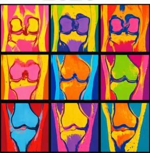
It is not a pristine tutorial dataset. Competitors will work from carefully pooled knee MRI images and their reports in 10 languages, sourced from 19 sites across 5 continents: the hedges, imperfect descriptions, and occasional errors that make clinical data both difficult and familiar.
The task is also not accuracy at all costs. Models must be efficient, and they must address a representative set of 12 knee abnormalities, not one model for one diagnosis.
That tension is the point. Building useful AI means confronting what radiology actually produces, then deciding what is worth measuring and improving.
As of August 17, there have been 12,853 joined users, 1,890 competitors across 1,761 active teams, and 11,456 submissions; the public leaderboard’s top, median, and last-listed scores are 0.951, 0.8745, and 0.468, respectively.
I am grateful to the RSNA staff, the Challenge Task Force, and especially Dr. Errol Colak for their leadership. I am equally thankful to my co-lead, Dr. Naveen Subhas. This Challenge could not have been pulled together by just one team: we owe this work to over 100 radiologists, biostatisticians, and data scientists across the world who volunteered their time reviewing reports, annotating images, offering advice, and contributing data.
We hope this challenge gives learners a practical place to begin and a clearer sense of the questions that remain.