Tag Archives: Radiology

FDA AI Guidance and the Hard Part of Transparency

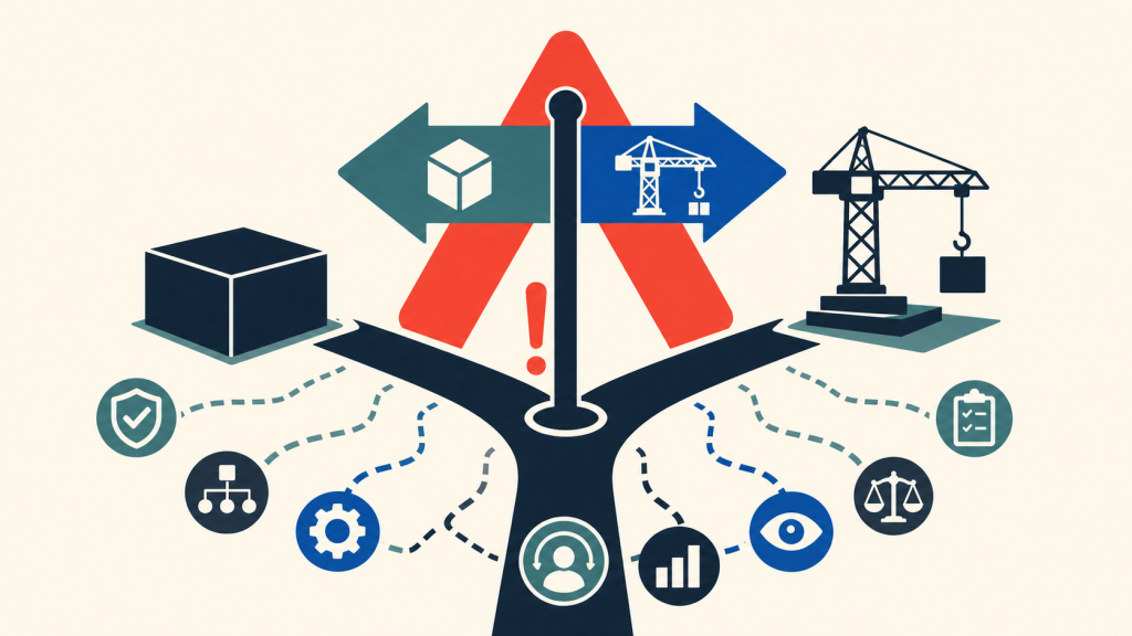
The least glamorous part of AI in radiology may turn out to be the most important: telling people what changed.

That sounds simple. It is not. A diagnostic AI tool may be trained on one dataset, validated on another, deployed inside a PACS or reporting workflow, monitored after release, and then updated when the model, threshold, input, interface, or intended environment changes. Somewhere in that chain, a radiologist is expected to decide whether to trust a box, a score, a contour, a triage flag, or a sentence.

Continue reading

AI Readiness as an Educational Obligation

I wrote a guest editorial in Academic Radiology about AI readiness as a practical educational obligation, rather than an optional informatics side quest.

The piece responds to a study of medical undergraduates and radiology trainees in China, but the issue is broader: enthusiasm and exposure do not equal competence. Radiologists need enough AI literacy to recognize workflow fit, automation bias, governance gaps, and the limits of machine suggestions. Most trainees will not become model developers.

They still need to become safe, skeptical operators who can use AI with confidence and accountability in real clinical environments, worldwide, across diverse resource settings today.

What Radiology Teaches Us About AI and the Workforce

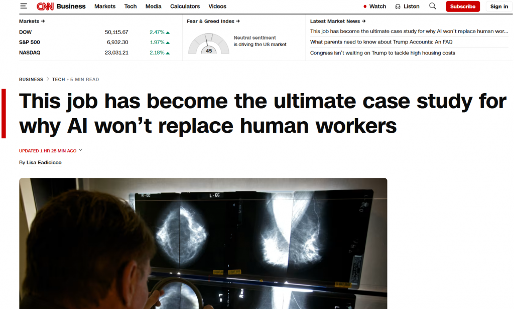
Health systems frequently ask whether artificial intelligence will ultimately reduce the need for clinicians. Radiology provides one of the clearest real-world answers to date. AI changes the mechanics of work far more than it changes the need for expertise.

I had the privilege to speak with CNN on this topic recently. The story is out now.

In imaging operations, AI is already being used to support exam prioritization, improve image reconstruction, and reduce friction in routine workflows. These contributions are meaningful, particularly in high-volume environments. But they do not displace clinical judgment, professional accountability, or responsibility for patient outcomes. In practice, the performance gains attributed to AI are inseparable from expert oversight and careful integration into clinical teams.

We use AI as a capacity and quality multiplier, not as a substitute for our training. That line is difficult to walk and gets thinner as products improve, but the thought matters. Deploying it primarily as a justification for workforce reduction or skill substitution introduces avoidable risk to patient safety and physician trust.

Success ultimately go to those investing in workflow, governance structures, and building solution that define clear roles for both humans and machines. Radiology may be an early example, but the underlying lesson extends well beyond imaging.

The FDA has an Idea, or 10, on Good Machine Learning Practice

Good machine learning practices goes beyond the chip

The U.S. FDA, Health Canada, and the UK’s MHRA have unveiled 10 guiding principles for Good Machine Learning Practice (GMLP) in developing AI/ML medical devices. These principles aim to ensure safety, efficacy, and quality in healthcare innovation. Key focuses include leveraging multi-disciplinary expertise, implementing good software and security practices, ensuring representative clinical study participants and data sets, maintaining independence between training and test data sets, and emphasizing the performance of the human-AI team. These guidelines also highlight the importance of clear user information, robust testing, and ongoing monitoring of deployed models to manage re-training risks and maintain performance.

Read the full GMLP draft on the FDA website.

1-Minute Summary

Here are the ten principles of GMLP.

Continue reading

AI+Human Better than Human in Neurodegenerative Imaging

Recent research underscores a leap in neuroimaging accuracy for Alzheimer’s disease diagnosis, emphasizing the superior performance of AI-assisted radiologists over either AI or humans alone. This collaborative approach marries the meticulous precision of AI with the nuanced understanding of human experts, potentially setting a new standard in the detection of amyloid-related imaging abnormalities. Specifically, it demonstrated superior performance in detecting amyloid-related imaging abnormalities (ARIA), crucial for amyloid-β–directed antibody therapy. This synergy enhances diagnostic precision and underscores the potential of AI-enhanced radiological diagnostics to improve patient care significantly.

How will this synergy between AI and human intelligence redefine the future of medical diagnostics? Can this model be the blueprint for addressing other complex diseases? This breakthrough prompts us to envision a healthcare landscape where technology and human expertise converge to offer unparalleled patient care.

Detailed study can be found in JAMA Network Open.

Your Origin Story for Data Science

There’s an origin story for every superhero; even those without superpowers (like Batman – that’s right) got started somewhere. What we sometimes forget is that there is also an origin story for every regular person, every profession, every hobby.

Source: Wikimedia Commons


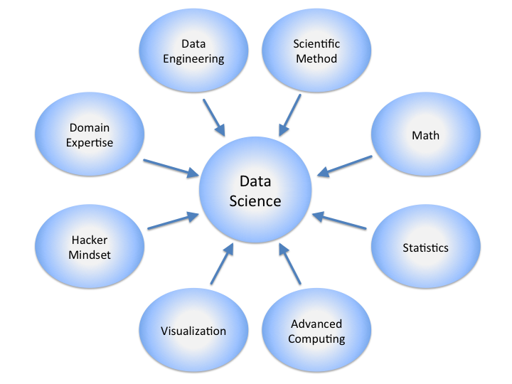
If you’re a radiologist looking to learn a few things in radiology data science, a simple web search will reveal a seemingly overwhelming amount of material you might have to know.

Fortunately, only a very small subset is necessary to start being productive.  Here are a few resources I used to get started.

Continue reading

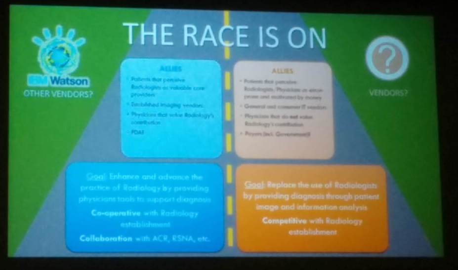
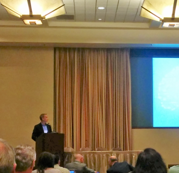
The [machine learning] race is on – Don Dennison

Continue reading

Machine learning, real opportunites: Dr. Keith Dreyer’s keynote sets tone for ISC 2016

Dr. Keith Dreyer opens with a keynote during the Intersociety Summer Conference (ISC) with description of data science and overview of how machine learning have evolved over time.

He describes that machines and humans inherently see things differently. Humans are excellent at object classification, recognition of faces, understanding language, driving, and imaging diagnostics. Continue reading

Five way to keep up coding skills when you are a full time radiologist

Radiologists have a day job (or a night job, depending on your precise definition of “radiologist.”) Many people want to learn the syntax of a computer language, while some want to keep up on existing skills.

If your goals are similar to mine, these might help.  Now these are not ways to learn to write code (I’ll write about that later), but ways to brush up on existing skills.

Here are five things to help keeping up your coding skills:

Work on a Project

Most radiology practices can be improved by better use of technology  Continue reading

Think Outside the (View) Box

Twenty years ago, medicine and surgery rounds used to start in the reading room.  Sitting in a dark room with a viewbox and an alternator, a senior radiologist greeted visiting clinical teams every day and reviewed their patients’ films.

With the advent of digitization and picture archive and communication system (PACS), the last 20 years saw a rapid evolution of radiology.  We read studies faster than ever, and radiology workflow focused extensively on the interpretation of images and the associated diagnostic report.

Recently, there has been a revival patient-centered care and communication.  Communication is the new radiology workflow.

I had the pleasure of writing about the importance of communication in radiology in a previous post. Just this month, a group at Beth Israel Deaconess Medical Center writes in American Journal of Roentgenology that despite our focus on critical value communication, the bulk (52%) of errors in radiology communication actually occur outside of results.

While most communication errors did not cause patient harm, 37.9% did affect patient care.  The radiology value chain, of course, begins as early as the decision to image and extends well into appropriate follow-up imaging of identified lesions (Enzmann, Radiology 2012).

Maybe it’s time we as radiologists take ownership of the whole imaging process, from the decision to image all the way to follow-up.

[Tweet “New AJR paper finds a majority of communication errors in radiology occur outside of results notification.”]